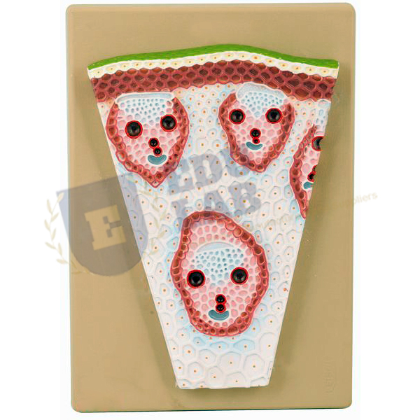
Animal Cell Model Manufacturer,Supplier and Exporter in India
Product Code : EL-BLE-10511
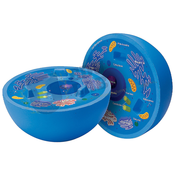
Quick Answer: The Animal Cell Model sets out the fine structure of an animal cell in bright colours, from the nucleus and mitochondria to the Golgi apparatus, centrioles and lysosomes, in one piece on a stand.
Last updated: October 2026
Key Features
Nucleus, endoplasmic reticulum, mitochondria, ribosomes and polysomes, and Golgi apparatus.
Centrioles, lysosomes and fat vacuoles also shown.
Extrusion of a Golgi vesicle and signs of pinocytosis show cell dynamics.
Brightly coloured components bring out how segmented the cell is.
One piece, on a stand with base.
Numbered, with key card.
Technical Specifications
Specification
Value
Construction
One piece
Mounting
Stand with base
Labelling
Numbered, with key card
Finish
Brightly coloured components
About This Product
Teachers use the model to name the organelles of an animal cell and point each one out with the numbered key. It also illustrates the cell at work, with a Golgi vesicle being released and signs of pinocytosis. The Animal Cell Model can be dispatched from India to Eswatini, Saint Vincent and the Grenadines, Grenada, Trinidad and Tobago, Morocco and Rwanda.
Frequently Asked Questions
Which organelles does the Animal Cell Model show?
The nucleus, endoplasmic reticulum, mitochondria, ribosomes and polysomes, Golgi apparatus, centrioles, lysosomes and fat vacuoles are all shown, numbered against a key card.
Does it show any cell processes?
Yes. It presents the extrusion of a Golgi vesicle and signs of pinocytosis to show cell dynamics.